The Ultimate Guide to Acing Your Anatomy Lab Practical Exam

One of the most challenging and important aspects of studying anatomy is the lab practical exam. This examination assesses a student’s ability to apply their knowledge of anatomy in a practical setting, such as identifying anatomical structures on preserved specimens or radiographic images. The lab practical exam is a crucial component of medical, dental, and other healthcare professional programs, as it allows students to demonstrate their understanding of anatomical structures and their relationships.
During the lab practical exam, students are typically presented with a series of stations or stations where they are expected to identify various anatomical structures. These structures may include bones, muscles, blood vessels, nerves, organs, and other tissues. The exam may also require students to describe the function or significance of these structures, as well as their relationship to other anatomical structures. In some cases, students may even be asked to perform specific tasks or procedures on the specimens.
Preparing for the anatomy lab practical exam requires extensive study and hands-on practice. Students must familiarize themselves with the anatomical structures they will be tested on, including their names, locations, and functions. They should also be able to identify these structures on different types of imaging, such as X-rays or CT scans. In addition to studying textbooks and lecture notes, students often find it helpful to use anatomical models, flashcards, and online resources to enhance their understanding.
Anatomy Lab Practical Exam

The Anatomy Lab Practical Exam is an essential part of any anatomy course. It is a comprehensive assessment that tests students’ knowledge and understanding of anatomical structures and their functions. During the exam, students are required to identify and locate various anatomical structures, such as bones, muscles, organs, and blood vessels. They are also expected to demonstrate their understanding of the relationships between these structures and their overall function within the human body.
To prepare for the Anatomy Lab Practical Exam, students must spend time studying and familiarizing themselves with the anatomical models and specimens in the lab. This includes learning the names, locations, and functions of different anatomical structures, as well as understanding the different systems of the human body, such as the skeletal, muscular, circulatory, and nervous systems. It is also important for students to develop good observation skills and attention to detail, as the exam often requires identifying and distinguishing between similar structures.
During the exam, students may be asked to perform a variety of tasks, such as labeling diagrams, identifying structures on specimens or models, and answering questions about the functions and relationships of different anatomical structures. Some exams may also include practical demonstrations, where students are required to perform specific tasks, such as locating specific structures on a cadaver or performing dissections to reveal hidden anatomical structures.
In order to succeed in the Anatomy Lab Practical Exam, students must not only possess a strong knowledge base but also be able to apply that knowledge in a practical setting. This requires critical thinking and problem-solving skills, as well as the ability to relate anatomical structures to their functions and understand how they work together to maintain the overall health and functioning of the human body.
Tips for Studying for an Anatomy Lab Practical Exam

Preparing for an anatomy lab practical exam can be challenging, as it requires a combination of theoretical knowledge and practical application. Here are some tips to help you effectively study and perform well on your exam.
1. Review the lab manual: Start by thoroughly reviewing the lab manual and familiarizing yourself with the different lab activities and structures you have studied. Pay attention to the relevant diagrams, terminology, and procedures mentioned.
Creating Flashcards
2. Create flashcards: To reinforce your learning, create flashcards for different anatomical structures, both visual and informational. Include key information such as functions, location, and specific anatomical features on the flashcards. Use these flashcards to quiz yourself regularly.
3. Practice hands-on: Take advantage of any available lab time to practice hands-on with anatomical models, cadavers, or virtual dissection tools. This will enhance your understanding and familiarity with the structures, making it easier to identify and recall them during the practical exam.
Study Groups and Labeling Diagrams

4. Join a study group: Collaborating with peers can be beneficial in anatomy studies. Engage in discussions, ask questions, and participate in group activities such as labeling diagrams or practicing identification of structures. This shared learning experience can help reinforce the material and foster a deeper understanding of anatomical concepts.
5. Label diagrams: A useful technique for studying is to label anatomical diagrams. Use clear, concise labeling to identify various structures, including bones, muscles, organs, and blood vessels. Regularly reviewing these labeled diagrams will improve your visual memory and aid in quick identification during the exam.
Practice Past Exams
6. Practice past exams: Obtain past lab practical exams, if available, and practice answering the questions within the time constraint. This will give you an idea of the format, type of questions asked, and the level of detail expected. By practicing under exam conditions, you can familiarize yourself with the pressure and work on creating efficient strategies for answering questions effectively.
7. Review classroom notes and lectures: While the focus of the practical exam is on the lab activities, it’s essential to review your classroom notes and lectures as well. This will provide you with a comprehensive understanding of the anatomical concepts, allowing you to apply your knowledge effectively during the exam.
By following these tips and maintaining a consistent study routine, you can improve your chances of performing well on your anatomy lab practical exam. Remember to stay organized, manage your time effectively, and seek clarification whenever needed. Good luck!
Lab Equipment and Supplies
The anatomy lab is equipped with a wide range of equipment and supplies that are essential for conducting practical exams and hands-on learning experiences. These tools allow students to explore the intricacies of human anatomy and develop their understanding of different anatomical structures.
Microscopes: Microscopes are one of the most fundamental tools in the anatomy lab. They enable students to closely examine slides of tissues, cells, and organs, allowing for a detailed analysis of their structure and function.
Dissection kits: Dissection kits include a variety of tools such as scalpels, dissecting scissors, forceps, and probes. These instruments are used for carefully dissecting cadavers or animal specimens, allowing students to study the anatomy in a hands-on manner.
Models and charts: The lab also contains a collection of anatomical models and charts that provide visual representations of various organ systems and structures. These models help students visualize complex anatomical concepts and enhance their understanding of the human body.
Preserved specimens: Preserved specimens, such as organs and body parts, are essential for studying anatomy. They allow students to observe the structure and characteristics of different tissues and organs, facilitating a deeper understanding of their functions.
Gloves and lab coats: Proper personal protective equipment, like gloves and lab coats, are essential for maintaining hygiene and safety in the anatomy lab. They protect students from potential hazards and ensure a clean and controlled environment for conducting experiments and dissections.
Measuring instruments: Measuring instruments like calipers and rulers are used to accurately measure and document the size and dimensions of anatomical structures. This is important for conducting precise observations and comparisons.
In conclusion, the anatomy lab is equipped with a wide range of tools and supplies that aid in the study of human anatomy. From microscopes to dissection kits, these resources enable students to explore and understand the complex structures and functions of the human body.
Exam Format and Structure

The Anatomy lab practical exam is an important component of the course that assesses students’ knowledge and understanding of human anatomy. The exam format consists of a series of stations, each focusing on a specific anatomical structure or system.
At each station, students are required to identify and label anatomical structures, such as bones, muscles, and organs. They may also be asked to demonstrate their knowledge of physiological processes or answer questions related to clinical applications of anatomy. The exam is typically timed, with students rotating through the stations within a specified time frame.
Station Format

Each station is set up with relevant anatomical models, specimens, diagrams, or images that students can refer to. They may be provided with a list of structures to identify or label, or they may be asked to identify structures independently. The stations are designed to test students’ ability to apply their knowledge of anatomy to real-life scenarios and clinical situations.
Students are expected to demonstrate their understanding of anatomical relationships, functions, and clinical significance. They may be asked to describe the function and location of a specific structure, identify any pathologies or abnormalities, or explain how a particular structure is involved in a specific physiological process. The exam requires students to think critically, make connections, and demonstrate their mastery of anatomical concepts.
Scoring and Grading
Each station is typically worth a certain number of points, and students are awarded points based on their performance at each station. The final score is calculated by adding up the points earned at each station. The grading scale may vary depending on the instructor or institution, but generally, a higher score indicates a better understanding of the subject matter.
It’s important for students to prepare for the exam by reviewing lecture notes, textbook material, and anatomical diagrams. Practice quizzes, flashcards, and group study sessions can also be helpful in reinforcing concepts and improving memorization. By familiarizing themselves with the format and structure of the exam, students can approach the Anatomy lab practical exam with confidence and perform to the best of their abilities.
Commonly Tested Topics

In an anatomy lab practical exam, there are several commonly tested topics that students should be familiar with. These topics cover various systems and structures of the human body that are typically studied in a lab setting.
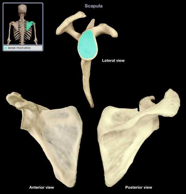
Skeletal System: One of the most important topics that is often tested in an anatomy lab practical is the skeletal system. Students are usually asked to identify and label different bones, such as the femur, humerus, and scapula. It is also common to be tested on the different types of joints and their movements.
- Identification of major bones
- Understanding of joint types and their movements
- Recognition of bone landmarks and features
Muscular System: Another commonly tested topic is the muscular system. Students may be asked to identify and name different muscles, such as the biceps brachii and quadriceps femoris. Knowledge of muscle attachments and actions is also important for this portion of the exam.
- Identification of major muscles
- Understanding of muscle attachments and actions
- Recognition of muscle groups and their functions
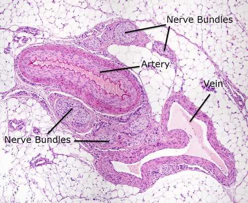
Cardiovascular System: The cardiovascular system is also a frequently tested topic. Students may be required to identify and label different structures of the heart, such as the atria, ventricles, and valves. Understanding the blood flow through the heart and major blood vessels is crucial for this part of the exam.
- Identification of heart structures
- Understanding of blood flow through the heart and major blood vessels
- Recognition of different types of blood vessels
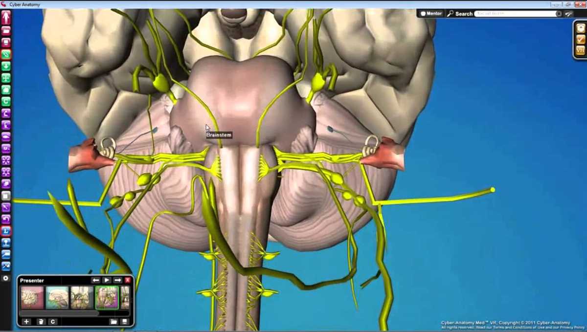
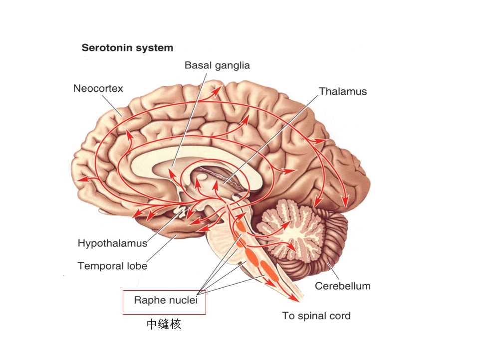
Nervous System: The nervous system is another important area that is commonly tested. Students may be asked to identify and name different parts of the brain, such as the cerebrum, cerebellum, and brainstem. Knowledge of the spinal cord, peripheral nerves, and their functions may also be tested.
- Identification of different parts of the brain and spinal cord
- Understanding of the functions of different parts of the brain
- Recognition of major peripheral nerves
Respiratory System: Lastly, the respiratory system is often tested in an anatomy lab practical exam. Students may be required to identify and label different structures of the respiratory system, such as the trachea, bronchi, and alveoli. Understanding the process of respiration and the role of the diaphragm is also important for this portion of the exam.
- Identification of respiratory structures
- Understanding of the process of respiration
- Recognition of the role of the diaphragm in breathing
Strategies for Success
When it comes to preparing for your anatomy lab practical exam, having a solid strategy in place can make all the difference. Here are some key tips to help you succeed:
- Study regularly: Rather than cramming all your studying into a few days before the exam, make it a habit to review your notes and materials regularly. This will help reinforce your knowledge and make the information more accessible during the exam.
- Use visual aids: Anatomy is a visual science, so utilizing visual aids such as diagrams, models, and interactive apps can greatly enhance your understanding. Creating your own visual aids, such as flashcards or mind maps, can also be a great way to reinforce key concepts.
- Practice with lab specimens: Take advantage of any opportunity to practice with actual anatomical specimens in the lab. Familiarize yourself with the different structures and functions by visually identifying them and understanding their relationships.
- Group study: Collaborating with classmates can be a valuable way to exchange knowledge and fill in any gaps in your understanding. Working together can also make the learning process more engaging and interactive.
- Seek help when needed: If you come across a topic or concept that you’re struggling with, don’t hesitate to seek help from your instructor, lab partners, or online resources. Getting a different perspective or explanation can often clarify things and make them easier to remember.
- Prepare for the practical aspect: In addition to understanding the theory, it’s crucial to practice the hands-on aspects of the exam. Familiarize yourself with the tools, techniques, and procedures you’ll be using, and make sure you’re comfortable with them before the exam.
- Stay motivated and manage stress: Anatomy can be challenging, but maintaining a positive attitude and finding ways to manage stress will help keep you motivated and focused. Take breaks when needed, stay organized, and practice self-care to help reduce anxiety and perform at your best.
By implementing these strategies, you can approach your anatomy lab practical exam with confidence and increase your chances of success. Remember, consistent effort and a well-rounded approach will go a long way in mastering this complex subject.