The Complete Solution: Gel Electrophoresis Worksheet Answer Key Uncovered

Gel electrophoresis is a widely used laboratory technique in molecular biology that separates and analyzes molecules based on their size and charge. It is a crucial tool for scientists studying DNA, RNA, and proteins, allowing them to gain valuable insights into a variety of biological processes. In order to interpret the results of gel electrophoresis experiments, it is important to have a clear understanding of the principles and techniques involved.
One way to assess students’ understanding of gel electrophoresis is through the use of worksheets. These worksheets typically include a series of questions and exercises that require students to analyze and interpret gel electrophoresis results. However, without an answer key, it can be difficult for students to determine if they have correctly completed the worksheet questions.
Having a gel electrophoresis worksheet answer key is essential for both students and educators. It provides students with a valuable resource for self-assessment, allowing them to check their answers and identify any areas of confusion or misunderstanding. For educators, the answer key serves as a guide for grading and provides a reference for explaining the correct answers to students.
Gel Electrophoresis Worksheet Answer Key
Gel electrophoresis is a powerful molecular biology technique used to separate and analyze DNA, RNA, or proteins based on their size and charge. This process involves placing the molecules in a gel matrix and applying an electric field, causing them to migrate through the gel. The gel acts as a sieve, with smaller molecules moving faster and farther than larger molecules.
The gel electrophoresis worksheet answer key provides the solutions and explanations for the questions and problems posed in a gel electrophoresis worksheet. This answer key can be used by students to check their answers and understand the concepts behind gel electrophoresis.
Example Questions and Answers:
1. What is the purpose of the loading dye in gel electrophoresis?
- The loading dye helps to track the movement of the sample during electrophoresis.
- It contains a colored dye that allows scientists to visually monitor the progress of the gel electrophoresis.
- Additionally, the loading dye contains a dense buffer that helps the sample settle into the wells of the gel.
2. How does gel electrophoresis separate molecules?
Gel electrophoresis separates molecules based on their size and charge. The gel matrix acts as a molecular sieve, with smaller molecules passing through the pores more easily than larger molecules. Additionally, the molecules are exposed to an electric field, causing them to migrate through the gel at different rates. Smaller molecules move faster and farther than larger molecules.
3. How can gel electrophoresis be used in DNA analysis?
- Gel electrophoresis can be used to determine the size of DNA fragments.
- DNA samples are loaded into wells in the gel and subjected to an electric field, causing the DNA fragments to migrate through the gel.
- The distance the fragments travel is inversely proportional to their size, allowing scientists to determine the size of the DNA fragments.
This gel electrophoresis worksheet answer key provides a valuable resource for students to check their understanding of gel electrophoresis concepts and ensure they are on the right track.
Overview of Gel Electrophoresis

Gel electrophoresis is a common laboratory technique used to separate and analyze DNA, RNA, and proteins. It involves the use of an electric field to move charged molecules through a gel matrix, allowing for their separation based on size, charge, or both.
The gel used in electrophoresis is typically made of agarose or polyacrylamide, and it is poured into a plastic or glass plate with wells at one end. The samples to be analyzed are loaded into these wells, and an electric current is applied across the gel. The molecules in the samples migrate through the gel at different rates depending on their size and charge, with smaller or more negatively charged molecules traveling faster than larger or positively charged ones.
The separated molecules can be visualized by staining the gel with dyes or using fluorescent tags. Different molecular weight markers are often used as a reference to estimate the size of the molecules being analyzed. The resulting banding patterns or separation profiles can provide valuable information about the composition, size distribution, and quantity of the molecules in the sample.
Gel electrophoresis has a wide range of applications in various fields, including molecular biology, genetics, forensics, and biotechnology. It is commonly used in DNA fingerprinting, gene expression analysis, protein analysis, and DNA sequencing. The technique is relatively simple, cost-effective, and versatile, making it an essential tool in many biological and biochemical research laboratories.
Principles of Gel Electrophoresis
Gel electrophoresis is a powerful technique used in the field of molecular biology to separate and analyze DNA, RNA, and proteins based on their size and charge. It is commonly used in research laboratories and forensic analysis to study genetic variations, identify specific DNA fragments, and analyze gene expression patterns. Gel electrophoresis utilizes an electric field to move charged molecules through a porous gel matrix, allowing for the separation of molecules based on their mobility.
To perform gel electrophoresis, a gel is prepared by mixing a polymer, typically agarose or polyacrylamide, with a buffer solution. Agarose gels are commonly used for larger DNA fragments, while polyacrylamide gels are used for separating smaller DNA fragments and proteins. The gel is poured into a gel tray or cassette and allowed to solidify. Wells are then created in the gel, providing spaces for loading the samples.
During the electrophoresis process, an electric field is applied across the gel, usually by connecting electrodes at each end. The samples, along with a loading buffer containing tracking dyes, are loaded into the wells. The electric field causes the charged molecules to migrate through the gel according to their size and charge. Smaller molecules will migrate faster and travel further through the gel, while larger molecules will migrate slower and remain closer to the well. This separation allows researchers to visualize and analyze the distribution of the molecules.
After the electrophoresis is complete, the gel is stained with a dye, such as ethidium bromide, that binds to the nucleic acids or proteins, making them visible under UV light. The bands formed on the gel represent the separated molecules, with each band corresponding to a specific DNA fragment or protein. These bands can be quantified and analyzed using specialized software to determine the size, quantity, and other characteristics of the molecules.
Gel electrophoresis has revolutionized molecular biology and has become an essential tool for genetic research and analysis. It provides valuable information about DNA, RNA, and protein molecules, allowing scientists to unravel the complexities of life at the molecular level.
Gel Electrophoresis Procedure
In gel electrophoresis, an electrical current is used to separate molecules based on their size and charge. This technique is commonly used in molecular biology to analyze DNA, RNA, and proteins. Gel electrophoresis can be performed using agarose or polyacrylamide gels.
To perform gel electrophoresis, first, the gel matrix is prepared. For agarose gels, agarose powder is dissolved in a buffer and heated until the powder is completely dissolved. The gel solution is then poured into a gel mold and allowed to solidify. For polyacrylamide gels, a crosslinking agent, such as ammonium persulfate, is added to a mixture of acrylamide and a bis-acrylamide solution. The gel solution is then poured into a gel mold and allowed to polymerize.
The next step is to prepare the samples that will be loaded onto the gel. The samples can be DNA, RNA, or protein samples, and they are usually mixed with a loading dye that contains a tracking dye and a denaturing agent. The loading dye helps visualize the movement of the samples during electrophoresis and allows for easy loading of the samples into the gel wells.
Once the gel is ready and the samples are prepared, the gel is placed in an electrophoresis tank filled with electrophoresis buffer. The gel is submerged in the buffer, and electrodes are connected to the tank. One electrode is placed at the top of the gel, and the other at the bottom. When the electrical current is applied, the molecules in the samples migrate through the gel matrix towards the positive electrode. The smaller molecules move faster and travel further, while larger molecules move slower and don’t travel as far.
After the electrophoresis run is complete, the gel is stained with a dye, such as ethidium bromide, that binds to DNA and RNA, or Coomassie brilliant blue, that binds to proteins. The stained gel is then visualized under UV light or by using a gel documentation system. The separated molecules appear as distinct bands on the gel, allowing researchers to analyze and compare the size and quantity of the molecules.
Gel electrophoresis is a powerful tool in molecular biology research and has various applications, including DNA fingerprinting, gene expression analysis, and protein purification. It allows scientists to separate and analyze molecules quickly, accurately, and efficiently.
Interpretation of Gel Electrophoresis Results
Gel electrophoresis is a powerful technique used to separate and analyze DNA molecules based on their size and charge. By applying an electric field to a gel matrix, DNA fragments can be separated and visualized as distinct bands on the gel. These bands provide valuable information about the DNA sample and can be used to answer various research questions.
Interpreting the results of gel electrophoresis involves several key steps:
- Size determination: The first step is to estimate the size of the DNA fragments in the sample. This can be done by comparing the migration distance of the fragments on the gel to a known standard ladder, which contains DNA fragments of known sizes. By plotting the migration distance of the DNA fragments on a standard curve, the approximate size of the unknown fragments can be determined.
- Banding patterns: The banding pattern on the gel provides information about the distribution of DNA fragments in the sample. A clear and distinct band indicates a single DNA fragment, while multiple bands suggest the presence of different-sized fragments. The intensity of the bands can also be informative, with stronger bands indicating a higher concentration of DNA.
- Comparative analysis: Gel electrophoresis results can be compared between different samples to identify similarities and differences. By running multiple samples simultaneously on the same gel, it is possible to determine if they contain the same DNA fragments or if there are variations in fragment size or abundance.
Overall, gel electrophoresis results provide valuable insights into the composition and characteristics of DNA samples. They can help in identifying DNA fragments of interest, analyzing genetic mutations, studying genetic diversity, and much more. The interpretation of these results requires careful analysis and comparison to known standards and other samples to draw meaningful conclusions.
Common Mistakes and Troubleshooting Tips
When performing gel electrophoresis, there are a few common mistakes that can occur. By being aware of these mistakes and knowing how to troubleshoot them, you can improve your gel electrophoresis results and ensure accurate analysis of your DNA or protein samples. Here are some common mistakes and their troubleshooting tips:
1. Inadequate loading buffer volume:
One common mistake is not adding enough loading buffer to the samples before loading them onto the gel. Loading buffer helps to visualize and track the movement of the samples during electrophoresis. If the loading buffer volume is insufficient, the samples may not migrate properly or may appear distorted on the gel. To troubleshoot this issue, make sure to add an appropriate volume of loading buffer to each sample, following the recommended guidelines provided.
2. Incorrect voltage or running time:
Another mistake that can affect gel electrophoresis results is using an incorrect voltage or running the gel for the wrong amount of time. Running the gel at too high or too low a voltage can cause the samples to migrate too quickly or too slowly, leading to inaccurate band sizes. Similarly, running the gel for too short or too long a time can result in incomplete separation of the sample bands. To troubleshoot this issue, double-check the recommended voltage and running time for your specific gel and adjust accordingly.
3. Contamination or degradation of samples:
Contamination or degradation of DNA or protein samples can also impact the gel electrophoresis results. Contaminants such as RNases or proteases can degrade the samples, causing smearing or faint bands on the gel. To avoid this, make sure to handle the samples with clean gloves and keep all reagents and equipment sterile. Additionally, store samples properly and avoid repeated freeze-thaw cycles. If contamination or degradation is suspected, consider repeating the experiment with fresh samples.
4. Uneven gel loading or sample handling:
Uneven gel loading or improper handling of samples can lead to uneven migration of bands on the gel. This can result in unclear or distorted band patterns. To troubleshoot this issue, make sure to load the samples evenly and at a consistent volume. Avoid touching the wells with the pipette tip while loading to prevent any disturbance in the sample distribution. Similarly, handle the gel carefully to avoid any smearing or distortion of the bands.
By being aware of these common mistakes and following the troubleshooting tips provided, you can optimize your gel electrophoresis experiments and obtain reliable and accurate results.
Applications of Gel Electrophoresis in Research and Medicine
Gel electrophoresis is a powerful technique that has found widespread applications in various fields, including research and medicine. It is a reliable and efficient method for separating and analyzing DNA, RNA, and proteins. The ability to separate molecules based on their size and charge has revolutionized the field of molecular biology and has led to numerous advancements in research and medical diagnostics.
Genetic research:

Gel electrophoresis is extensively used in genetic research to analyze and compare DNA fragments. Researchers can obtain valuable information about genetic variation, mutation, and inheritance patterns by studying the size and distribution of DNA fragments on a gel. This allows them to study genetic disorders, identify potential disease-causing mutations, and understand the underlying genetic mechanisms of various diseases.
Forensic analysis:
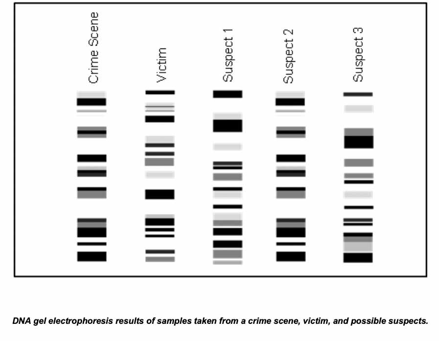
Gel electrophoresis plays a crucial role in forensic analysis, particularly in DNA profiling. By running DNA samples from crime scenes alongside DNA samples from suspects, scientists can determine whether the suspect’s DNA matches that found at the crime scene. This technique has been instrumental in solving criminal cases and ensuring justice.
Medical diagnostics:

Gel electrophoresis is widely used in medical diagnostics for the detection and identification of various diseases. For example, it is used to detect genetic disorders, infectious diseases, and cancer biomarkers. By analyzing specific DNA or protein markers on a gel, medical professionals can identify the presence of disease and monitor its progression. This information is vital for accurate diagnosis, prognosis, and treatment planning.
Quality control:
Gel electrophoresis is also used in quality control processes, especially in the pharmaceutical and biotechnology industries. It allows manufacturers to ensure the integrity and purity of their products by verifying the presence and quantity of specific molecules. This ensures the safety and effectiveness of drugs, vaccines, and other biotechnological products.
Conclusion:
Gel electrophoresis is an indispensable tool in research and medicine. Its ability to separate and analyze molecules based on size and charge has revolutionized the field of molecular biology and has numerous applications. From genetic research to forensic analysis and medical diagnostics, gel electrophoresis is a cornerstone technique that continues to contribute to scientific advancements and improve human health.